Get the latest article directly in your inbox. Sign up today


Insight by Oticon Medical - MRI exams

Download article


By: Oticon Medical
Date: 8 July 2019

Magnetic Resonance Imaging (MRI) is a well-established technique used in clinical settings around the world to provide professionals with valuable diagnostic and prognostic information about a patient’s health. Users of the Oticon Medical Neuro Zti can be reassured that their implant will not stop them undergoing this type of scan. 


MRI and CI patients

MRI scans are an established diagnostic tool used for body, as well as brain scans, ranging in strength from 0.2 Tesla to 7 Tesla. Recent estimates show that around 79% of MRI scanners in use around the world are 1.5 Tesla, making this the most common type by far1.


The Neuro Zti implant is fully compatible with 1.5 Tesla scans with the magnet in place for both body and head scans. The implant has been designed to ensure this type of scan is safe and painless and does not produce ‘magnet flip’ as the magnet is held firmly in place by the rigid implant structure2. This means it can’t dislodge and remains locked in even after multiple MRI exams, while the implant itself remains stable throughout the MRI scan thanks to a unique screw fixation system that securely attaches it to the bone. This has been confirmed by an independent study from Berlin3. Furthermore, if needed, the implant structure of the Neuro Zti is designed for easy magnet removal.


Why radiologists often want the magnet removed regardless of Tesla strength

Brain scans account for 20-25% of MRI scans among people worldwide1,4,5. For CI patients, this figure increases to 49%6, making brain scans the most common MRI exams they are likely to undergo. For this reason, it’s important to understand how an MRI scan affects a cochlear implant – and also how the images are affected by the magnet.


Leaving the implant magnets in place during head scans of either 1.5 Tesla or 3 Tesla can usually influence the image quality. The magnet may create artefacts that distort the result and prevent reliable diagnosis. This is even more common in scans of 3 Tesla because of the additional magnetic strength. See figure 1.

  • 620x620-mri-f1

    Figure 1

    Example of  MR image of head at 1.5T(left) and at 3T (right) with magnet in situ7.

  • 620x620-mri-f2

    Figure 2

    Example of MR image of head at 1.5T (left) and 3T (right) after magnet removal with non-magnetic spacer in situ7.

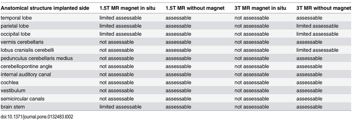
In fact, when asked radiologists stated that they could not make an accurate diagnosis from images scanned with 3 Tesla with the magnets in place because of the interference created by the magnets7 (see table below). This can be clearly seen on figure 1 of the MRI head scan, where the magnet creates artefacts. When this happens, the magnet has to be removed. For this reason, its ease of removal is an important factor when considering an implant.


Summary of the visibility of the rated anatomical structures at the side of the cochlea implant; with and without magnet at 1.5T and 3T.

1150x400-summary 

Ref: Wagner, F., Wimmer, W., Leidolt, L., Vischer, M., Weder, S., Wiest, R., Mantokoudis, G. and Caversaccio, M.D., 2015. Significant artifact reduction at 1.5 T and 3T MRI by the use of a cochlear implant with removable magnet: an experimental human cadaver study. PloS one, 10(7), p.e0132483.


Neuro Zti – direct, easy and quick magnet removal

The Neuro Zti is 3 Tesla compatible without magnet and is designed for easy magnet removal under local anesthesia with only a small skin incision.  The implant is attached using screws and not drilled into the skull. With the dedicated Oticon Medical surgical tool, the magnet is removed in a few steps and can then be replaced again after the scan. 


620x175-remove-magnet
The dedicated surgical tool simplifies magnet removal 


Furthermore, the Neuro Zti implant magnet can be removed multiple times without damaging the body of the implant. This can be an issue for CI magnets cased in silicone that can loosen over time causing the magnet to flip and migrate8. For younger cochlear implant patients who are likely to undergo several MRI scans during their lifetime, ease of removal should be part of the decision making process when choosing an implant system.


1. The Royal College of Radiologists, the College of Radiographers and the Institute of Physics and Engineering in Medicine (2017). Magnetic resonance imaging (MRI) equipment, operations and planning in the NHS - Report from the Clinical Imaging Board. London, UK. Retrieved from www.rcr.ac.uk/sites/default/files/cib_mri_equipment_report.pdf

2. Oticon Medical CI Unique – January 2019 (the Oticon Medical Uniue logo Indicates an Oticon Medical unique technology or accessory in the CI industry at the time of this publication)

3. Todt, I., Rademacher, G., Grupe G., Stratmann A., Ernst, A., Mutze S., Mittmann P. (2018). Cochlear implants and 1.5 T MRI scans: the effect of diametrically bipolar magnets and screw fixation on pain. Journal of Otolaryngology - Head and Neck Surgery (2018) 47:11

4. www.ameli.fr/fileadmin/user_upload/documents/Pratiques_et_recommandations_sur_l_IRM.pdf   

5. Saadat S. et al, Overuse or underuse of MRI scanners in private radiology centers in Tehran, International Journal of Technology Assessment in Health Care 24(3):277-81 · February 2008

6. Grupe G, Wagner J, Hofmann S, Stratmann A, Mittmann P, Ernst A, Todt I. Prevalence and complications of MRI scans of cochlear implant patients: English version. HNO. 2017 Jan;65(Suppl 1):35-40. doi: 10.1007/s00106-016-0129-7. PMID: 26886493

7. Wagner, F., Wimmer, W., Leidolt, L., Vischer, M., Weder, S., Wiest, R., Mantokoudis, G. and Caversaccio, M.D., 2015. Significant artifact reduction at 1.5 T and 3T MRI by the use of a cochlear implant with removable magnet: an experimental human cadaver study. PloS one, 10(7), p. e0132483.

8. F. Hassepass1, V. Stabenau1, S. Arndt1, R. Beck1, S. Bulla2, T. Grauvogel1, A. Aschendorff: Magnet Dislocation: An Increasing and Serious Complication Following MRI in Patients with Cochlear Implant